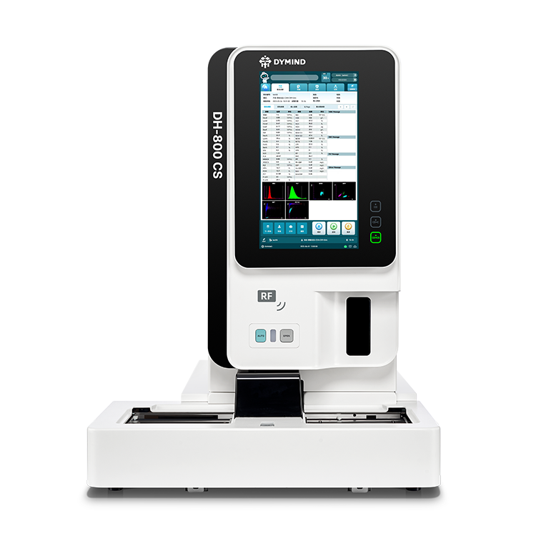
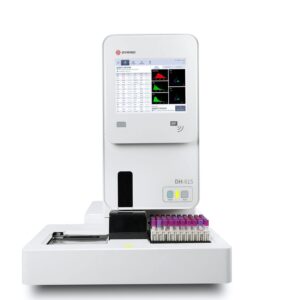
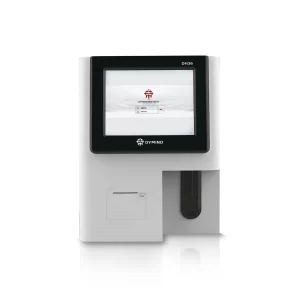
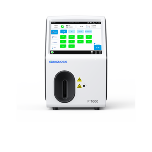
Automatic hematology analyzer is a device used clinically to analyze blood cells, differentiate the 5 white blood cell components, measure hemoglobin concentration and measure reticulocytes and red blood cells. enucleated (NRBC), and body fluids (cerebrospinal fluid, peritoneal fluid, pleural fluid, and synovial fluid).
Analysis modes: DH-800[T7]CS has 30 analysis modes including tests: CBC (complete blood count), RET (reticulocytes), PLT-F (platelets), DIFF ( white blood cell differentiation), CRP (C-reactive protein), SAA (Serum amyloid A),WPC (pristine blood cells).

Operating principle: The automatic hematology testing machine uses the sheath flow impedance method, laser scattering method, fluorescent dye method and scattering flow cell method. semiconductor laser-based flow cytometry to count and differentiate blood cells; Colorimetry is used to measure hemoglobin concentration and latex-enhanced immunonephelometry is used to measure the concentration of specific proteins.
Sheath-flow electrical impedance method principle: The analyzer applies the sheath-flow electrical impedance method to count and differentiate red blood cells (RBC) and platelets (PLT). The RBC and PLT sample are surrounded by a high-speed sheath solution flow, which in turn passes through the aperture and generates an electrical pulse according to the Coulter principle, the measurement module’s processing circuits amplify the electronic signal received and compared with the RBC/PLT classification voltage threshold, and counted the number of electrical pulses with amplitudes within the range of RBC and PLT. RBCs and PLTs are classified based on the voltage amplitude of the electrical pulses that are within the classification threshold, and the number of RBCs and PLTs are also counted based on the number of electrical pulses of each type. Furthermore, the electrical pulses classified as RBC and PLT are divided into equal intervals based on the amplitude of the electrical pulse, and the number of electrical pulses in each interval is counted to calculate the RBC cellularity parameter. and PLT.
Principle of fluorescence dyeing method: The analyzer applies fluorescence dyeing method to process pre-analytical samples for DIFF, WNR, WPC (according to device customization), RET and PLT-F tests. During the reaction of DIFF, WNR and WPC, lysate is first added to lyse specific cells, leaving the cells intact for analysis and dyes are added to label and stain the nucleic acids in the cells. cell; while in the RET and PLT-F reactions, a dilute solution is added to make the cells spherical, and then a dye is added to label and stain the nucleic acids in the cells. The purity of the nucleic acid of different cell types, cells at different stages, and abnormal cells will be different, which in turn leads to different amounts of fluorescent dye. When a laser scattering flow cell is performed, the laser light emits fluorescence at a longer wavelength than the light incident upon irradiation of the fluorescent dye bound inside the cell, and the fluorescent light emitted The output is captured by a side fluorescent receiver, where the fluorescence intensity is proportional to the concentration of the bound dye. Pre-scattered light is proportional to the size of the spherical cell, side-scattered fluorescent light represents the amount of stained nucleic acids inside the cell, and side-scattered light represents the complexity of the cell, from Then multiple scatter plots can be generated to count and differentiate cell types.
Principle of semiconductor laser scattering flow cell method: The analyzer adopts semiconductor laser scattering flow cell method to perform analysis of DIFF, WNR, WPC (according to device customization), RET and PLT-F after the sample has been pre-processed for analysis by fluorescence staining. A certain amount of blood cells are stained and labeled with a fluorescent dye, and injected into a conical flow chamber containing a dilute solution at a certain rate through a nozzle inside the flow chamber. The blood cells are enveloped by a high-speed flow of sheath solution arranged in rows and one by one passing through the center of the flow chamber, and irradiated by a laser at a certain wavelength at next to the flow chamber. Light is scattered at different angles as cells pass through the laser, stimulating fluorescent dyes bound inside the cells to emit fluorescent light at longer wavelengths. Three optical probes in different directions receive front-scatter, side-fluorescence and side-scatter signals and convert them into electrical pulse signals. Processing circuits amplify these electrical pulses, and they are used to plot 3D scatterplots of blood cell size, information within the cell, and fluorescence levels. In which, forward scatter (FSC) shows the difference in blood cell size, side scatter (SSC) shows the complexity of particles inside blood cells, and side fluorescence shows the level of staining of blood cells. cell.
Principle of colorimetric method: The analyzer applies colorimetric method to measure HGB concentration. In the reaction tank, lysate is added to the diluted sample and red blood cells are lysed to release hemoglobin. Hemoglobin combines with sodium dodecyl sulfate in the lysate to form a hemoglobin complex. The above solution is transferred to the colorimetric tank for analysis. The LED emits a beam of light at a certain wavelength on one side of the colorimetric tank, and irradiates the hemoglobin complex solution. The emitted light is measured by an optical probe on the opposite side and converted into an electronic signal, which becomes a voltage signal after being amplified and filtered by the electronic circuit. Based on the Beer-Lambert Law, the hemoglobin concentration of the sample is calculated by comparing the measured voltage with the voltage generated by the measured background light when no sample has been added to the colorimetric bath (when only the solution is present). diluent in the tank).
Principle of latex scattering immunoturbidimetric method: The analyzer applies immunoturbidimetric method to measure the concentration of specific proteins such as CRP and SAA. The analyzer emits parallel monochromatic light at a certain wavelength into the reaction tank. Light is scattered when it encounters the antibody-antigen complex. The analyzer captures the scattered light and converts the light intensity into a voltage signal and amplifies it. The specific protein concentration of a sample is calculated by comparing the measured voltage with the voltage generated by background light measured before adding the sample to the reaction tank. The intensity of scattered light is directly proportional to the number of complexes, meaning that the more complexes, the stronger the scattered light because the number of antigens increases.
Specifications:
| Size | 655mm (W) x 867mm (D) x 868mm (H)
115kg |
| Operating conditions | Temperature: 15°C-32°C
Humidity: 30-85% Pressure: 70-106 kPa |
| Power supply | Voltage: AC 100V-240V (±10%)
Frequency: 50Hz/60Hz(±1 Hz) Power: 660VA |
| Wattage | Up to 110 samples/hour |
| Sample type | Whole blood, capillary blood, diluted blood, body fluids. |
| Parameter | 44 reporting parameters (whole blood), 7 parameters (body fluids) + 152 research parameters (whole blood) and 11 research parameters (body fluids) |
| Screen | 12.1 inch touch screen |
| Communicate | USB, LAN, and HL7 ports with bidirectional LIS |


