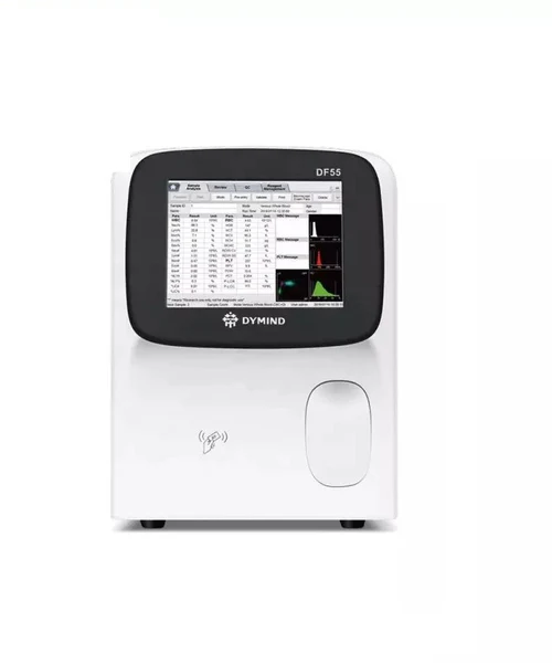
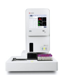
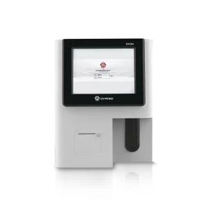
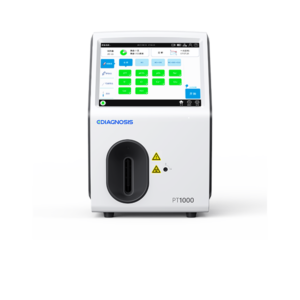
The automatic hematology testing machine is a device used clinically to analyze blood cells, differentiate the 5 components of white blood cells, and measure hemoglobin concentration.
Analysis mode: CBC (complete blood count), CBC+DIFF (complete blood count + white blood cell differential).
Operating principle: Hematology testing machine uses electrical impedance method to analyze RBC and PLT data; colorimetric method to measure HGB concentration; and laser-based flow cytometry to analyze WBC data.
Laser scattering flow cytometry method principle: After a certain amount of blood is aspirated and diluted with a certain amount of reagent, it is injected into the flow chamber. Surrounded by the dilute solution, the blood cells are passed through the center of the flow chamber in a column at high speed, and passed through a laser beam that causes the light to scatter at many different angles. The intensity of the scattered light represents the size of the blood cells and the intracellular density. In particular, narrow-angle scattered light signals represent cell size, while medium-angle and wide-angle scattered light signals represent intracellular information (information about the nucleus and cytoplasm). . Optical probes receive these scattered information and convert them into electrical pulse signals. The electrical pulse data was then used to plot a 2D scatter plot.
Principle of colorimetric method: WBC/HGB diluent is introduced into the HGB tank and mixed with a certain amount of lysate, converting hemoglobin into a complex that can be measured at 525 nm. An LED is mounted on one side of the tank and emits a monochromatic beam of light at a wavelength of 525 nm. Light passes through the sample and is measured by an optical probe on the opposite side. The signal is then amplified and the voltage is measured and compared to a blank sample (sample measured when there is only dilute solution in the tank).
Electrical impedance method principle: This method works by measuring the change in resistance created by a blood cell as it passes through an aperture of known size. The electrode is placed at both ends of the solution on both sides of the aperture to form an electrically conductive path. As each cell passes through the aperture it creates a change in the resistance between the two electrodes. This change produces an electrical pulse that can be measured. The number of electrical pulses generated is equal to the number of cells passing through the aperture, and the amplitude of each electrical pulse is proportional to the volume of the corresponding cell. Each electrical pulse is amplified and compared to the internal reference voltage channel, and only electrical pulses with sufficiently high amplitude are accepted. If the electrical pulse generated has an amplitude higher than the lower threshold value of WBC/BAS/RBC/PLT, it is considered WBC/BAS/RBC/PLT.
Specifications:
| Size | 431mm (D) x 364mm (W) x 498mm (H)
28kg |
| Operating conditions | Temperature: 15°C-30°C
Humidity: 20-90% Pressure: 70-106 kPa |
| Power supply | Voltage: AC 100V-240V (±10%)
Frequency: 50Hz/60Hz(±1 Hz) Power: ≤220VA |
| Wattage | Up to 60 samples/hour |
| Sample type | Whole blood, capillary blood, diluted samples |
| Parameter | 31 parameters, including 25 reporting parameters |
| Screen | 10.4 inch touch screen |
| Communicate | Bi-directional USB, LAN, and LIS ports |


